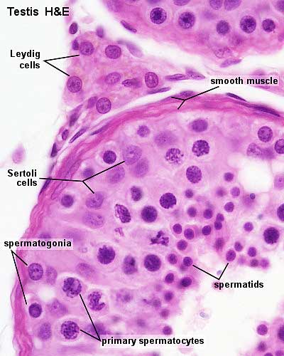
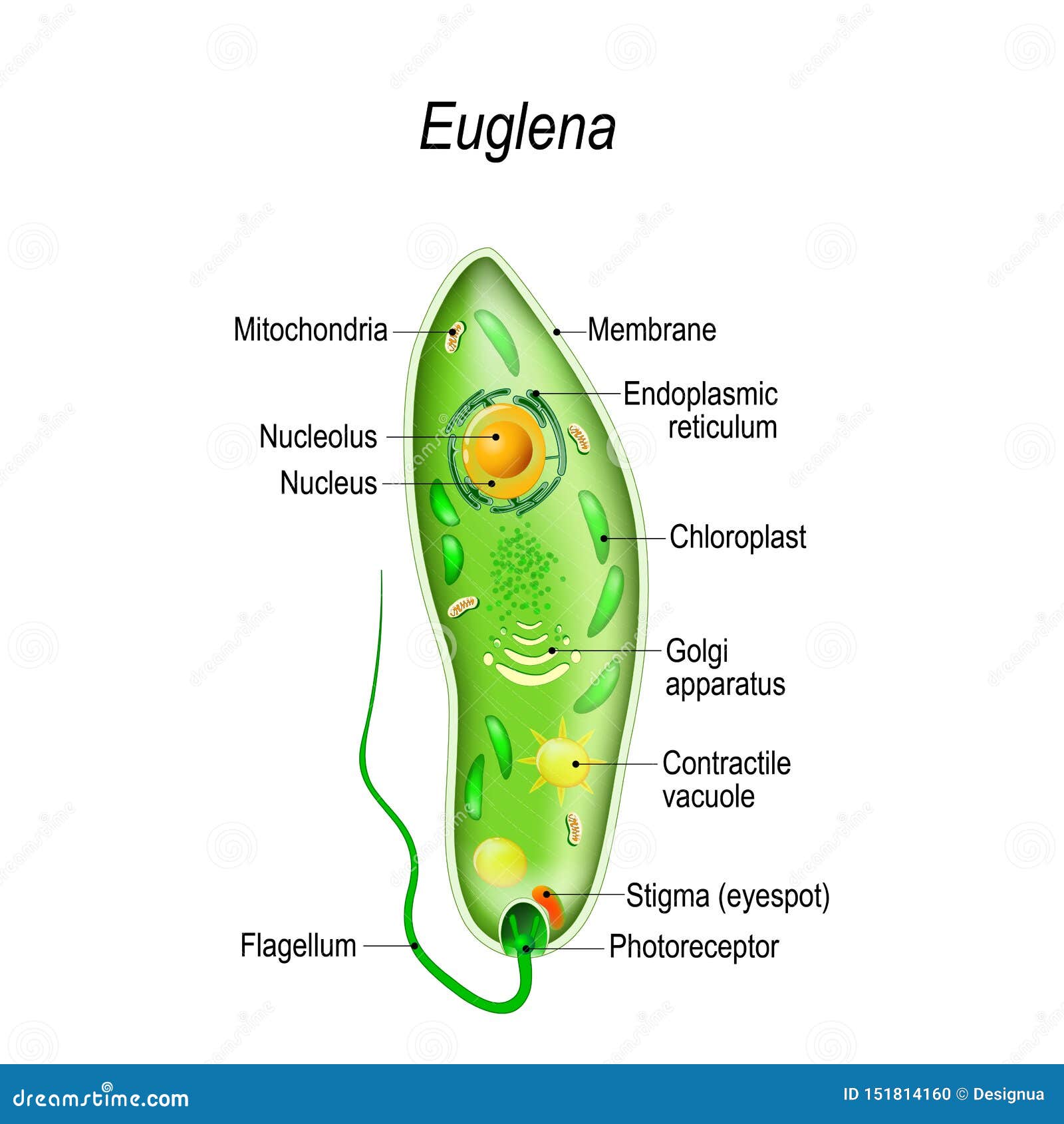
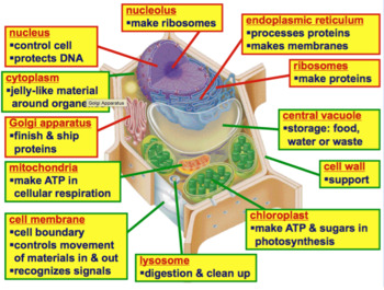
40 cell diagram and labels
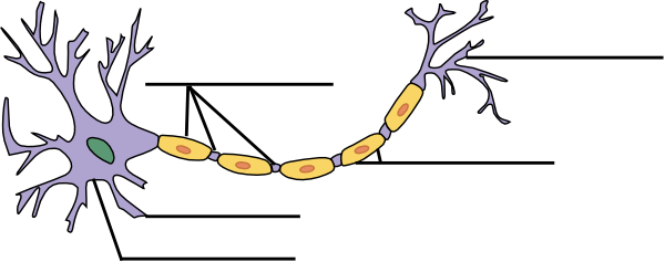
NERVOUS SYSTEM WORKSHEET - St. Francis Preparatory School 1. The diagram below is of a nerve cell or neuron. Add the following labels to the diagram: Axon Myelin sheath Cell body Dendrites Muscle fibers Axon terminals 2. Color in the diagram as suggested below. Axon - purple Axon Terminals – orange Myelin sheath – yellow Cell body – blue Dendrites – green Muscle fibers - red 3. Help Online - Origin Help - Adding Unicode and ANSI ... Adding ANSI Characters to Text Labels. These are older methods which pre-date Origin 2018 and Unicode support. If you are entering text in the Object Properties dialog box, in worksheet column cells, or you are typing prefixes or suffixes in dialog box text boxes (such as you see on the Tick Labels page of the Axis dialog box), you can access the ANSI character set using this procedure:
CELL MEMBRANE LABEL Diagram | Quizlet Practice labeling the parts of the cell membrane Learn with flashcards, games, and more — for free. Home. Subjects. Explanations. ... identifies or labels the cell. Receptor protein. receives information. Heads. part of the phospholipid that loves water (hydrophili) - points to the most outside and inside of cell ... Animal Cell Diagram. 14 ...

Cell diagram and labels
Chord diagram – from Data to Viz A chord diagram represents flows or connections between several entities (called nodes). Each entity is represented by a fragment on the outer part of the circular layout . Then, arcs are drawn between each entities. Plant and Animal Cell: Labeled Diagram, Structure ... Both plant and animal cells have similar types of architecture. They are made up of cell boundaries, cytoplasm, nucleus and several cellular organelles. Structure. Description and function. Cell Wall. 1. Non-living, rigid, outer boundary. 2. Made up of cellulose, hemicellulose, pectin, lignin, etc. Cell: Structure and Functions (With Diagram) Eukaryotic Cells: 1. Eukaryotes are sophisticated cells with a well defined nucleus and cell organelles. 2. The cells are comparatively larger in size (10-100 μm). 3. Unicellular to multicellular in nature and evolved ~1 billion years ago. 4. The cell membrane is semipermeable and flexible. 5. These cells reproduce both asexually and sexually.
Cell diagram and labels. PDF Human Cell Diagram, Parts, Pictures, Structure and Functions One of the few cells in the human body that lacks almost all organelles are the red blood cells. The main organelles are as follows : cell membrane endoplasmic reticulum Golgi apparatus lysosomes mitochondria nucleus perioxisomes microfilaments and microtubules 2 A Labeled Diagram of the Animal Cell and its Organelles ... A Labeled Diagram of the Animal Cell and its Organelles There are two types of cells - Prokaryotic and Eucaryotic. Eukaryotic cells are larger, more complex, and have evolved more recently than prokaryotes. Where, prokaryotes are just bacteria and archaea, eukaryotes are literally everything else. The Cell Cycle Coloring Worksheet - WPMU DEV Jul 10, 2017 · 7. During what phase of the cell cycle does the cell grow? 8. During what phase of the cell cycle does the cell prepare for mitosis? 9. How many stages are there in mitosis? 10. Put the following stages of mitosis in order: anaphase, prophase, metaphase, and telophase. 11. Put the following stages of the cell cycle in order: G2, S, G1, M. 12. 03 Label the Cell Diagram - Quizlet Cell Biology 03 Label the Cell STUDY Learn Flashcards Write Spell Test PLAY Match Gravity Created by muskopf1TEACHER Terms in this set (14) Nucleus Control center of the cell Nucleolus Ribosome synthesis Rough Endoplasmic Reticulum Protein transport Smooth Endoplasmic Reticulum Lipid synthesis Mitochondrion Cellular Respiratoin Golgi Apparatus
Plant Cell Diagram Labeled 6th Grade Simple : Functions ... Plant Cell Diagram Labeled 6th Grade. Here, let's study the plant cell in detail. Plant cell parts are almost similar to animal cells with few exceptions and functional differences. We all remember that the human body is very problematic and a method I found out to understand it is via the way of human anatomy diagrams. Cell Membrane Diagram Labeled : Functions and Diagram Cell Membrane Diagram Labeled Monday, March 22nd 2021. | Diagram Cell Membrane Diagram. There are no organelles in the prokaryotic cells, i.e., they have no internal membrane systems. While lipids help to give membranes their flexibility, proteins monitor and maintain. Labeled Plant Cell With Diagrams - Science Trends The parts of a plant cell include the cell wall, the cell membrane, the cytoskeleton or cytoplasm, the nucleus, the Golgi body, the mitochondria, the peroxisome's, the vacuoles, ribosomes, and the endoplasmic reticulum. Parts Of A Plant Cell The Cell Wall Let's start from the outside and work our way inwards. Plant Cell - Definition, Structure, Function, Diagram & Types Plant Cell Structure. Just like different organs within the body, plant cell structure includes various components known as cell organelles that perform different functions to sustain itself. These organelles include: Cell Wall. It is a rigid layer which is composed of cellulose, glycoproteins, lignin, pectin and hemicellulose.
Unit 1 Biology and Disease Cell structure & function Practice ... cell between points P and Q. Give your answer in um. Show your working. Answer . . um (2 marks) (a) (a) (i) (ii) Name organelle Y. (1 mark) There are large numbers of organelle Y in this cell. Explain how these organelles help the cell to absorb the products of digestion. (2 marks) The diagram shows an epithelial cell from the small intestine. Plant Cell Diagram - Science Trends A plant cell diagram, like the one above, shows each part of the plant cell including the chloroplast, cell wall, plasma membrane, nucleus, mitochondria, ribosomes, etc. A plant cell diagram is a great way to learn the different components of the cell for your upcoming exam. Plants are able to do something animals can't: photosynthesize. Animal Cell Diagram with Label and Explanation: Cell ... Diagram of Animal Cell Below is the diagram of the animal cell which shows the organelles present in it. The cell is covered with cytoplasm which consists of cell organelles in it. The nucleus is covered with a rough Endoplasmic Reticulum and other organelles each designed for a specific purpose. A Well-labelled Diagram Of Animal Cell With Explanation The animal cell diagram is widely asked in Class 10 and 12 examinations and is beneficial to understand the structure and functions of an animal. A brief explanation of the different parts of an animal cell along with a well-labelled diagram is mentioned below for reference. Also Read Different between Plant Cell and Animal Cell
Plant Cells: Labelled Diagram, Definitions, and Structure The cell wall is made of cellulose and lignin, which are strong and tough compounds. Plant Cells Labelled Plastids and Chloroplasts Plants make their own food through photosynthesis. Plant cells have plastids, which animal cells don't. Plastids are organelles used to make and store needed compounds. Chloroplasts are the most important of plastids.
IXL | Plant cell diagrams: label parts | 8th grade science Improve your science knowledge with free questions in "Plant cell diagrams: label parts" and thousands of other science skills.
Neuron Cell Diagram With Labels - nervous tissue scientist ... Neuron Cell Diagram With Labels - 17 images - cognitive science 107a wk1d2 flashcards quizlet, nervous tissue and physiology week 11 flashcards easy, yfp neuron clip art at vector clip art online, neuronal cell types current biology,
Human Cell Diagram, Parts, Pictures, Structure and ... One of the few cells in the human body that lacks almost all organelles are the red blood cells. The main organelles are as follows : cell membrane endoplasmic reticulum Golgi apparatus lysosomes mitochondria nucleus perioxisomes microfilaments and microtubules Diagram of the human cell illustrating the different parts of the cell. Cell Membrane
Diagram Of Plant And Animal Cells To Label Teaching ... Student Objectives:Identify cell organelles on animal, plant, and bacteria cell diagrams.Describe the functions of major cell organelles.Analyze the similarities and differences among: a) eukaryotic versus prokaryotic cells b) plant versus animal cellsStudents will follow directions to label the major cell organelles in Animal, Plant, and Bacteria cells, then complete matching activities with ...
Animal Cell - Free printable to label + Color -kidCourses.com Can you label and color these important parts of the animal cell?. NUCLEUS control center for cell (cell growth, cell metabolism, cell reproduction). NUCLEOLUS synthesizes rRNA. RIBOSOMES the site of protein building, this is where translation takes place (mRNA in language of nucleic acids is translated into the language of amino acids). RER (Rough Endoplasmic Reticulum) synthesizes proteins ...
Plant Cell Diagram without Labels | Plant cells worksheet ... Plant Cell Structure. Plant Life Cycle Worksheet. Plant Cell Project. Plant Lessons. Free plant cell worksheets for students to identify and label the parts. Younger students can use our free plant cell coloring pages, while older students can learn the parts of a cell. snoopygirl11.
Label Cell Parts | Plant & Animal Cell Activity ... Student Instructions Create a cell diagram with each part of plant and animal cells labeled. Include descriptions of what each organelle does. Click "Start Assignment". Find diagrams of a plant and an animal cell in the Science tab. Using arrows and Textables, label each part of the cell and describe its function.
Nuclear envelope - Wikipedia Nesprin-mediated connections to the cytoskeleton contribute to nuclear positioning and to the cell’s mechanosensory function. KASH domain proteins of Nesprin-1 and -2 are part of a LINC complex (linker of nucleoskeleton and cytoskeleton) and can bind directly to cystoskeletal components, such as actin filaments , or can bind to proteins in ...
Cell Worksheets | Plant and Animal Cells Plant Cell Diagram | Animal Cell Diagram. Featured in this printable worksheet are the diagrams of the plant and animal cells with parts labeled vividly. This enhanced visual instructional tool assists in grasping and retaining the names of the cell parts like mitochondrion, vacuole, nucleus and more with ease.
Cell Organelles- Definition, Structure, Functions, Diagram Cilia and Flagella are tiny hair-like projections from the cell made of microtubules and covered by the plasma membrane. Structure of Cilia and Flagella Cilia are hair-like projections that have a 9+2 arrangement of microtubules with a radial pattern of 9 outer microtubule doublet that surrounds two singlet microtubules.
A Labeled Diagram of the Plant Cell and Functions of its ... A Labeled Diagram of the Plant Cell and Functions of its Organelles We are aware that all life stems from a single cell, and that the cell is the most basic unit of all living organisms. The cell being the smallest unit of life, is akin to a tiny room which houses several organs. Here, let's study the plant cell in detail...
Interactive Cell Model - CELLS alive Cell Wall. Chloroplast. Smooth Endoplasmic Reticulum. Rough Endoplasmic Reticulum. Ribosomes. Cytoskeleton. RETURN to CELL DIAGRAM ...
Coordination Chemistry III: Tanabe-Sugano Diagrams and Charge ... d7Tanabe-Sugano Diagram E / B ∆o/ B 4F 2G 2Eg 2T1g 2A1g 2T2g 4P 4A2g 4T1g (4P) 4T2g 4T1g (4F) small ∆o High Spin large ∆o Low Spin Complexes with d4-d7 electron counts are special •at small values of ∆o/B the diagram looks similar to the d2diagram •at larger values of ∆o/B, there is a break in the diagram leading to a new ground ...
Animal Cell Diagram | Animal cells worksheet, Cell diagram ... Description Teach your students all about the inner working of an animal cell with the help of this hand-drawn animal cell diagram. This pdf packet contains 6 versions of the diagram, to help you teach and also quiz your students: 1. Labeled Animal Cell Diagram - Color 2. Labeled Animal Cell Diagram - Black & White 3.
IXL | Animal cell diagrams: label parts | 7th grade science Improve your science knowledge with free questions in "Animal cell diagrams: label parts" and thousands of other science skills.
Cell: Structure and Functions (With Diagram) Eukaryotic Cells: 1. Eukaryotes are sophisticated cells with a well defined nucleus and cell organelles. 2. The cells are comparatively larger in size (10-100 μm). 3. Unicellular to multicellular in nature and evolved ~1 billion years ago. 4. The cell membrane is semipermeable and flexible. 5. These cells reproduce both asexually and sexually.
Plant and Animal Cell: Labeled Diagram, Structure ... Both plant and animal cells have similar types of architecture. They are made up of cell boundaries, cytoplasm, nucleus and several cellular organelles. Structure. Description and function. Cell Wall. 1. Non-living, rigid, outer boundary. 2. Made up of cellulose, hemicellulose, pectin, lignin, etc.


Post a Comment for "40 cell diagram and labels"