39 cell wall diagram with labels
Cell wall structure with plant cellular parts description outline diagram Yeast cell vector illustration. Labeled organism closeup structure diagram. Root hair cell collecting mineral nutrients and water from soil, biological labeled plant system diagram Fungi as basic fungal cell and multicellular fungus structure outline diagram Bacterial cell structure with anatomical inner parts sections outline diagram Label the Cell Membrane - Labelled diagram - Wordwall Label the Cell Membrane - Labelled diagram channel protein, cholesterol, external cell environment, hydrophilic (water loving) part of phospholipid bilayer, peripheral protein.
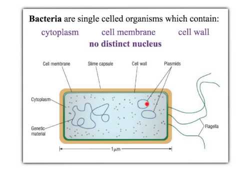
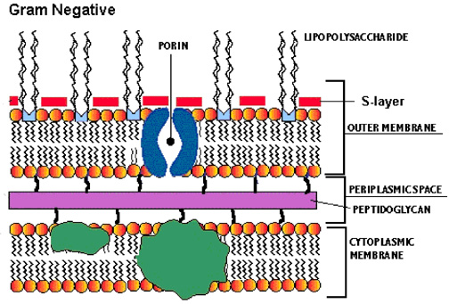
Bacteria in Microbiology - shapes, structure and diagram The bacteria shapes, structure, and labeled diagrams are discussed below. Sizes The sizes of bacteria cells that can infect human beings range from 0.1 to 10 micrometers. Some larger types of bacteria such as the rickettsias, mycoplasmas, and chlamydias have similar sizes as the largest types of viruses, the poxviruses.

Cell wall diagram with labels
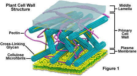
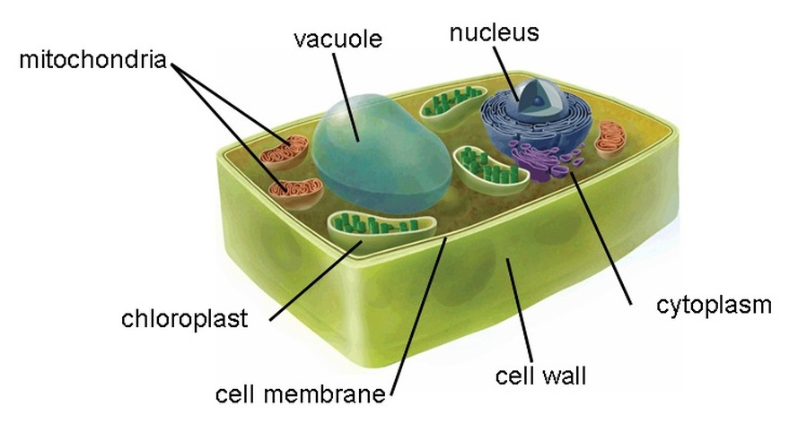
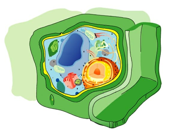
Plant Cell Diagram | Science Trends A plant cell diagram, like the one above, shows each part of the plant cell including the chloroplast, cell wall, plasma membrane, nucleus, mitochondria, ribosomes, etc. A plant cell diagram is a great way to learn the different components of the cell for your upcoming exam. Plants are able to do something animals can't: photosynthesize. Plant Cell Wall Stock Illustrations - Dreamstime Download 21,398 Plant Cell Wall Stock Illustrations, Vectors & Clipart for FREE or amazingly low rates! New users enjoy 60% OFF. 189,527,695 stock photos online. Stock Photos; ... Cell wall structure with plant cellular parts description outline diagram. Labeled educational model components description with hemicellulose, pectin and. Structure of Bacterial Cell (With Diagram) - Biology Discussion These are long filamentous, cytoplasmic appendages, 12-30 μm in length, protruding through the cell wall and contain contractile protein flagellin. These are organs of locomotion. Fimbriae or pili: These are thin, short filaments (0.1-1.5 μm x 4 to 8 nm) extruding from the cytoplasmic membrane, also called pili. They are made of protein (pilin).
Cell wall diagram with labels. Plant Cells: Labelled Diagram, Definitions, and Structure The cell wall is made of cellulose and lignin, which are strong and tough compounds. Plant Cells Labelled Plastids and Chloroplasts Plants make their own food through photosynthesis. Plant cells have plastids, which animal cells don't. Plastids are organelles used to make and store needed compounds. Chloroplasts are the most important of plastids. Learn the parts of a cell with diagrams and cell quizzes - Kenhub Two major regions can be found in a cell. The first is the cell nucleus, which houses DNA in the form of chromosomes. The second is the cytoplasm, a thick solution mainly comprised of water, salts, and proteins. The parts of a eukaryotic cell responsible for maintaining cell homeostasis, known as organelles, are located within the cytoplasm. Structure of Cell Wall (With Diagram) - Biology Discussion Cells with secondary wall consist of five layers a three layered secondary wall, the primary wall and the middle lamella. In some cells, such as primary xylem, the secondary thickening materials are laid down in such a way that various patterns are formed on the cell wall, e.g. annular, spiral, reticulate, scalariform and pitted. Human Cell Diagram, Parts, Pictures, Structure and Functions Diagram of the human cell illustrating the different parts of the cell. Cell Membrane. The cell membrane is the outer coating of the cell and contains the cytoplasm, substances within it and the organelle. It is a double-layered membrane composed of proteins and lipids. The lipid molecules on the outer and inner part (lipid bilayer) allow it to ...
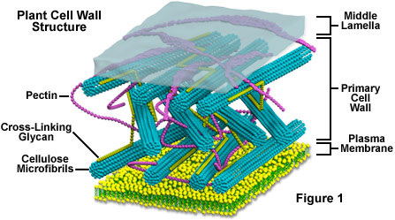
Labeled Plant Cell With Diagrams - Science Trends The parts of a plant cell include the cell wall, the cell membrane, the cytoskeleton or cytoplasm, the nucleus, the Golgi body, the mitochondria, the peroxisome's, the vacuoles, ribosomes, and the endoplasmic reticulum. Parts Of A Plant Cell The Cell Wall Let's start from the outside and work our way inwards. Cell Organelles- Definition, Structure, Functions, Diagram In a plant cell, the cell wall is made up of cellulose, hemicellulose, and proteins while in a fungal cell, it is composed of chitin. A cell wall is multilayered with a middle lamina, a primary cell wall, and a secondary cell wall. The middle lamina contains polysaccharides that provide adhesion and allow binding of the cells to one another. Plant and Animal Cell: Labeled Diagram, Structure, Function - Embibe Cell Wall: 1. Non-living, rigid, outer boundary. 2. Made up of cellulose, hemicellulose, pectin, lignin, etc. 3. There are many layers, like the middle layer, primary cell wall in a typical plant cell wall. 4. Fungal cell wall is made up of chitin (not cellulose). 5. Protective and provide shape and size. 6. Found only in plant cells. Plasma ... Elodea Leaf Cell Diagram Elodea Leaf Cell Diagram The Elodea leaf is composed of two layers of cells. Only one layer of cells is in focus when using the high. Examining elodea (pondweed) under a compound microscope. solution) and a coverslip and observe the chloroplasts (green structures) and the cell walls.
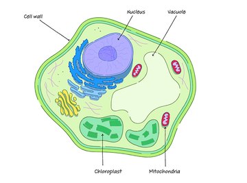
File:Plant cell wall diagram-en.svg - Wikimedia Commons Size of this PNG preview of this SVG file: 497 × 288 pixels. Other resolutions: 320 × 185 pixels | 640 × 371 pixels | 1,024 × 593 pixels | 1,280 × 742 pixels | 2,560 × 1,483 pixels. Original file (SVG file, nominally 497 × 288 pixels, file size: 57 KB) File information. Structured data. Captions. Plant cell label - Labelled diagram - Wordwall Nucleus , Cell membrane , Cytoplasm, Mitochondria, Vacuole, chloroplast, Cell wall. Plant cell label. Share Share by Mcintosh. Like. Edit Content. Embed. More. Leaderboard. Show more Show less . This leaderboard is currently private. Click Share to make it public. This leaderboard has been disabled by the resource owner. ... A Well-labelled Diagram Of Animal Cell With Explanation The animal cell diagram is widely asked in Class 10 and 12 examinations and is beneficial to understand the structure and functions of an animal. A brief explanation of the different parts of an animal cell along with a well-labelled diagram is mentioned below for reference. Also Read Different between Plant Cell and Animal Cell Animal Cell Labelling Activity | Primary Resources | Twinkl This Animal Cell Labelling Activity is the perfect way to help children consolidate their learning on the biology of cells. Cells are the building blocks of life, and they're important for all budding young biologists to understand. With that in mind, we have created a range of resources to help those who teach teach their students about cells:

Cell Wall - Components | Function of Cell Wall | Plant Cell Wall
animal cell with labels mitochondria cell mitochondrion risk disease cells study edu powerhouses shape heart began upends theories current virginia uva va mitochondrial dna. ARTimus Prime: 6th Grade- Watercolor Cells artimusprimecobra.blogspot.com. grade animal plant cell cells 6th diagram artimus prime. Cell plant label worksheets. This is the cell wall. it protects ...

Phage Therapy (Biocontrol*): Light and Shade Visit new site : Sellanophagetherapy.blogspot.com ...
03 Label the Cell Diagram | Quizlet Start studying 03 Label the Cell. Learn vocabulary, terms, and more with flashcards, games, and other study tools.
Cell Wall - Cell Organelles
Bacterial Cell Structure Labeling Diagram | Quizlet Cell Wall A semirigid casing that provides structural support and shape for the cell Cytoskeleton Long fibers of proteins that encircle the cell just inside the cytoplasmic membrane and contribute to the shape of the cell Pilus Appendage used for drawing another bacterium close in order to transfer DNA Glycocalyx
Cells - Rumney Marsh Academy Science Revere, Massachusetts
A Labeled Diagram of the Plant Cell and Functions of its Organelles A Labeled Diagram of the Plant Cell and Functions of its Organelles We are aware that all life stems from a single cell, and that the cell is the most basic unit of all living organisms. The cell being the smallest unit of life, is akin to a tiny room which houses several organs. Here, let's study the plant cell in detail...
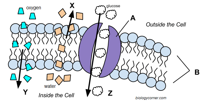
Cell Membrane and Transport
PDF Plant Cell Diagram - Edrawsoft Plant Cell Golgi vesicles Golgi apparatus Ribosome Smooth ER(no ribosomes) Nucleolus Nucleus Rough ER(endoplasmic reticulum) Large central vacuole Amyloplast(star ch grain) Cell wall Cell membrane Chloroplast Vacuole membrane Raphide crystal Mitochondrion Druse crystal
plant, animal, bacteria and yeast cells - Revision Cards in IGCSE Biology
Animal Cell Diagram with Label and Explanation: Cell ... - Collegedunia Diagram of Animal Cell Below is the diagram of the animal cell which shows the organelles present in it. The cell is covered with cytoplasm which consists of cell organelles in it. The nucleus is covered with a rough Endoplasmic Reticulum and other organelles each designed for a specific purpose.

Scheme of a cell with cell wall
Plant Cell: Diagram, Types and Functions - Embibe Exams Plant Cell Wall It is a rigid layer that is composed of cellulose, glycoproteins, lignin, pectin and hemicellulose. It is located outside the cell membrane and is completely permeable. The primary function of a plant cell wall is to protect the cell against mechanical stress and to provide a definite form and structure to the cell.
Proteobacteria - microbewiki
PDF Plant Anatomy: Images and diagrams to explain concepts A diagram of a prokaryotic cell. It lacks organelles and is much smaller and simpler. (LadyofHats Mariana Ruiz. Public Domain). PLANT ANATOMY AND PHYSIOLOGY: IMAGES AND DIAGRAMS TO EXPLAIN 7. 1.2 CELL WALL The cell wall is initially deposited on the surface of the middle lamella. This primary cell wall occurs on the surface of all plant cells ...

Labeled Diagram Of Cell Wall - Diagram Media
Label Cell Parts | Plant & Animal Cell Activity | StoryboardThat Click "Start Assignment". Find diagrams of a plant and an animal cell in the Science tab. Using arrows and Textables, label each part of the cell and describe its function. Color the text boxes to group them into organelles found in only animal cells, organelles found in only plant cells, and organelles found in both cell types.
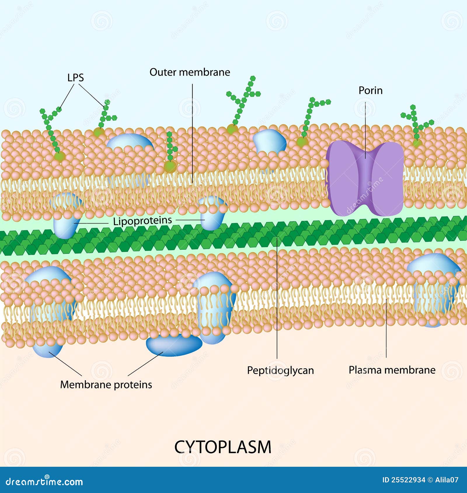
Gram Negative Bacterial Cell Wall Stock Images - Image: 25522934
Plant Cell Diagram The Label A diagram of a plant cell with the organelles labeled Nucleolus 8 . The cell wall is made of nonliving material called cellulose Learn about the similarities and differences between plant Both plant cells and animal cells are Eukaryotic cells Glossary of Plant Cell Anatomy Terms In plant cells, ATP is produced in the cristae of mitochondria and ...
Cell Wall - Definition, Function & Structure | Biology Dictionary
Structure of Bacterial Cell (With Diagram) - Biology Discussion These are long filamentous, cytoplasmic appendages, 12-30 μm in length, protruding through the cell wall and contain contractile protein flagellin. These are organs of locomotion. Fimbriae or pili: These are thin, short filaments (0.1-1.5 μm x 4 to 8 nm) extruding from the cytoplasmic membrane, also called pili. They are made of protein (pilin).
Picture
Plant Cell Wall Stock Illustrations - Dreamstime Download 21,398 Plant Cell Wall Stock Illustrations, Vectors & Clipart for FREE or amazingly low rates! New users enjoy 60% OFF. 189,527,695 stock photos online. Stock Photos; ... Cell wall structure with plant cellular parts description outline diagram. Labeled educational model components description with hemicellulose, pectin and.

Image result for illustrations of a cell and different components | Organelles, Eukaryotic cell ...
Plant Cell Diagram | Science Trends A plant cell diagram, like the one above, shows each part of the plant cell including the chloroplast, cell wall, plasma membrane, nucleus, mitochondria, ribosomes, etc. A plant cell diagram is a great way to learn the different components of the cell for your upcoming exam. Plants are able to do something animals can't: photosynthesize.

Cell Mitosis

What is a Cell Wall - 99Science
Cell Wall: Definition, Structure & Function (with Diagram) | Sciencing

The Life-Sized Cell Wall - Important Information
Post a Comment for "39 cell wall diagram with labels"